Investigating the brain - Higher
Modern science has allowed scientists to discover how different parts of the brain function. Neuroscientists have been able to map various regions of the brain to particular functions by studying patients with brain damage, electrically stimulating different parts of the brain and using Magnetic Resonance Imaging - A computer that can scan the human body using magnetic fields and radio waves, used in medicine. scanning techniques.
A well-documented example of brain damage is of Phineas Gage, who in 1848 had a serious accident whilst laying railway tracks and an iron rod went through his skull.
Phineas survived the accident and could still walk and talk, but it was documented that his personality changed following it. It was noted that he lost his inhibitions socially and emotionally.
Doctors realised the changes in Phineas were due to the damage in the particular parts of the brain that the iron rod had passed through. This important case allowed scientists to examine the effect of the injuries on his brain activity.
Non-invasive brain procedures include:
Electrical stimulation
Scientists have stimulated different parts of the brain with a weak electrical current and asked patients to describe what they experienced. If the motor area is stimulated, the patient makes an involuntary movement. If the visual area is stimulated, they may see a flash of colour. EEGs (Electroencephalograms) can be created and studied, to observe the electrical activity in the brain.
There are three modern techniques used to scan brains:
- MRI (magnetic resonance imaging)
- CT (computed tomography)
- PET (positron emission tomography)
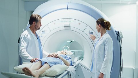
MRI scanners use strong magnetic fields and radio waves to show details of brain structure and function. Patients are asked to perform various tasks and, by looking at the scan, scientists can see which parts of the brain are active when the task is carried out. The patient lies on a bed which moves into the machine. Some people can feel claustrophobic inside an MRI scanner.
CT scanners are similar to MRIs. The patient lies on a bed which passes through a ring of equipment (not into the machine like in MRI). The ring takes a series of x-rays from different angles. These are processed by a computer to allow the doctors to see inside brains and other parts of the body.
PET scanners detect gamma rays that radiate from a chemical compound called a tracer. PET scans are used to detect high levels of metabolic reactions inside a person. Before going into the scanner the patient consumes the tracer. This travels to any area of the body which has unusually high levels of metabolic reactions. This is often a tumour and so PET scans are used to detect A disease caused by normal cells changing so that they grow and divide in an uncontrolled way. The uncontrolled growth causes a lump called a tumour to form..