Magnification
Equation
Magnification can be worked out from a photograph or drawing using the equation below:
The same unit of measurement should be used when making the calculation - metre (m), millimetre (mm) or micrometre (µm).
To convert millimetres into micrometres, multiply by 1000.
The above equation can be rearranged in order to calculate the actual length of the cell and the magnification used as well as the length of the image.
Actual Length = length of the Image divided by the Magnification.
Magnification = length of the Image divided by the Actual Length.
Scale bar
Magnification can be calculated using a scale bar. This is a line drawn near the photograph or drawing which has a label showing the actual length of the bar before being magnified.
Working out magnification:
- Measure the scale bar image (beside drawing) in mm.
- Convert to µm (multiply by 1000).
- Magnification = scale bar image divided by actual scale bar length (written on the scale bar).
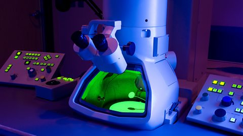
Electron microscopes
Continually increasing the magnification past a certain point does not increase the detail visible in an image.
The most important property of a microscope is its The fineness of detail that can be seen in an image - the higher the resolution of an image, the more detail it holds. In computing terms, resolution is measured in dots per inch (dpi). - the ability to show detail.
The best light microscope can show details that are 0.2µm apart and need a magnification of roughly x1500 so that our eyes can see it - this allows us to see larger cell structures.
Electron microscopes pass beams of electrons through a specimen and have a much greater resolution than a light microscope.
Their resolution can show details that are 0.0001µm apart - allowing us to clearly see inside the parts of a cell e.g. inside a Part of a cell that carries out photosynthesis. Chlorophyll inside the chloroplast traps sunlight. or Structures in the cytoplasm of all cells where aerobic respiration takes place (singular is mitochondrion)..